頭頂葉

(図1)前頭葉
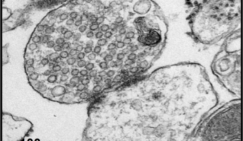
右図では、頭頂葉の神経細胞の形態を二光子顕微鏡でイメージングした。
頭頂葉
右図では、頭頂葉の神経細胞の形態を二光子顕微鏡でイメージングした。
日付:
2016年9月20日
Copyright OIST (Okinawa Institute of Science and Technology Graduate University, 沖縄科学技術大学院大学). Creative Commons Attribution 4.0 International License (CC BY 4.0).
タグ
Research


