


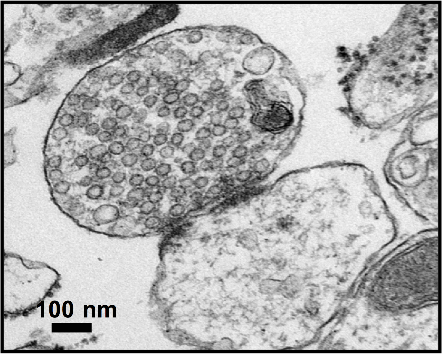
A high magnification image of synapse obtained by electron microscopy

Figure 3. A high magnification image of synapse obtained by electron microscopy (zoom x 20000 times).
The image shows an isolated synapse from a brain sample before mass spectrometry analyses. The pre-synapse typically shows many vesicles containing neurotransmitters kept attached to the post-synapse (Z. Taoufiq, OIST 2013).
Date:
29 August 2013
Copyright OIST (Okinawa Institute of Science and Technology Graduate University, 沖縄科学技術大学院大学). Creative Commons Attribution 4.0 International License (CC BY 4.0).
Tags
Research
Share on:


