To See OIST on a Grain of Salt
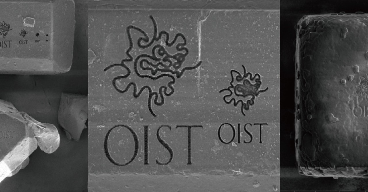
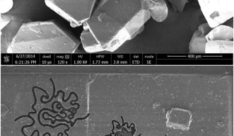
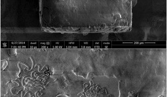
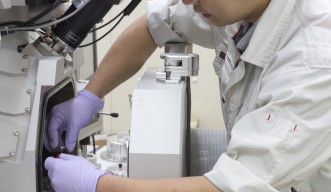
Imagine the Okinawa Institute of Science and Technology Graduate University logo. Now, imagine shrinking the logo so that it can fit onto a grain of salt. A few weeks ago, the Biology Resources Section carved the design onto an actual grain of salt, a grain of sugar, and a grain of rice. The section ran this project to create a learning tool for the OIST Visitor’s Center. They used the Focused Ion Beam/Scanning Electron Microscope, or FIB/SEM, a hybrid machine that images one surface of a sample, and then mills a thin layer off of that surface before imaging the next layer down.
At OIST, the FIB/SEM machine is often used to visualize biological samples, but it can be used to visualize samples from almost any field. First, the machine shines a beam of electrons at the sample. When the electrons strike the sample, they bounce back at different speeds and in different directions, depending on the material they strike. A detector measures the electrons bouncing off of the sample, and produces a microscopic image. Then, a focused beam of gallium ions mills the sample, removing a layer as thin as one micrometer. The machine repeats this process of imaging and milling until the entire sample is complete, producing a stack of images. Usually, a researcher uses a computer to leaf through the images and see how cells change throughout the biological sample, layer by layer.
The amount of material in the sample and the thickness of each layer determine how long this milling and imaging process will take. For example, one recent scan required about three weeks of constant processing to complete a sample about a bit smaller than one-third of the head of a pin. During this time, the FIB/SEM milled the sample 20 nanometers at a time, producing ten gigabytes of data and 1000 images, each a 40 micrometer square.
Toshio Sasaki, who works in the Biology Resources Section, decided that beyond imaging, he could create some unusual images with the FIB/SEM. He used the gallium ion beam to carve the OIST logo onto a grain of rice, a grain of salt, and a grain of sugar. Then he took photos of each one, each showing a scale to put the nanoscale logos into perspective.
“The salt was the most challenging,” said Sasaki, explaining that the imaging works best on samples that are neutral, or have no charge. Because the gallium beam has a positive charge and the electron beam has a negative charge, a sample with a charge repels either one beam or the other. Both sugar and salt are made of atoms with positive and negative charges, giving them conductivity. In order to make each sample non-conductive, or neutral, Sasaki coated each sample in a thin layer of gold, a non-conductive metal. However, salt’s particles have stronger charges than sugar, making it more difficult to neutralize the salt sample. For this reason, the logo on the salt looks a little messier than the one on the sugar.
The resulting images might not teach researchers anything new about cells, but they are useful for teaching visitors about microscopic imaging. The images will be on display in the OIST Visitors Center.
By Poncie Rutsch
Specialty
For press enquiries:
Press Inquiry Form


