New Unit Profile: Beautiful Viruses
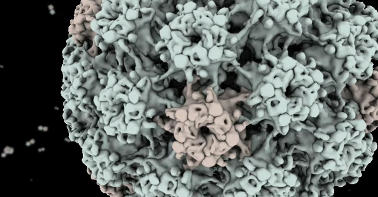
Matthias Wolf was supposed to be a pharmacist. He studied pharmacology as an undergraduate, even though he was “more interested in the essential properties of life,” he says. But then he changed tack, earning a PhD in biophysics and focusing his research on exploring the structures of biological molecules. Now, as the head of OIST’s new Molecular Cryo-Electron Microscopy Unit, his main muse is virus structure.
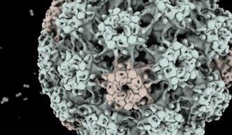
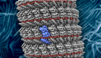
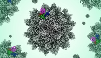
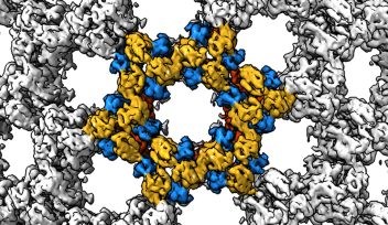
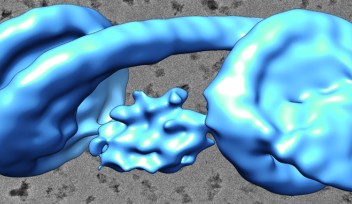
Virus coatings are symmetrical, which makes it easier to study their structures, Prof. Wolf explains. “They’re like the Death Star in Star Wars. To some people they look menacing; to me they look beautiful,” he says. To image viruses, Prof. Wolf and his team use electron microscopy, which bombards samples with high-energy electrons. Since electron bombardment can damage the structure of the proteins that make up a virus’s coat, he uses a low dose of electrons, and then makes up for the loss in image quality by combining information from many images of the same virus to get a fuller picture.
Using the papilloma and polyoma viruses as subjects, Prof. Wolf’s team is focusing on how viruses latch on to cells, poke holes in the cell membrane, and inject their genetic material into their hosts, thereby infecting them. “Our goal is to create a molecular movie that shows what happens,” Prof. Wolf says. To do that, they need vast amounts of data, which they plan to collect by automating OIST’s high-end electron microscope to take many images by itself. They also need to devise new computational methods to process all that data.
Though he left pharmacology behind years ago, Prof. Wolf’s work may ultimately bring him full circle: he hopes that by finding out exactly how viruses infect cells, he will identify potential drugs that would interfere with that mechanism. Thus, his appreciation of viruses’ beauty may help to lessen their menace.
Specialties
Research Unit
For press enquiries:
Press Inquiry Form


