


Cryogenic electro-microscopy photos

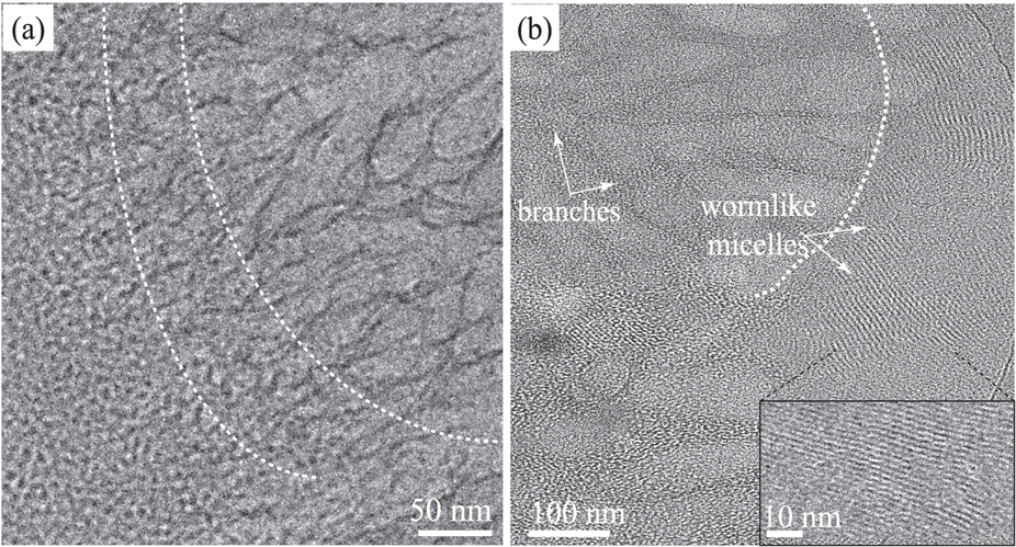
A) Cryogenic electro-microscopy photos captured the transitions (highlighted by the white dashed lines) from spherical micelles to wormlike micelles to branches structures (from left to right) at 15°C. B) Transition from wormlike micelles to branches.
A) Cryogenic electro-microscopy photos captured the transitions (highlighted by the white dashed lines) from spherical micelles to wormlike micelles to branches structures (from left to right) at 15°C. B) Transition from wormlike micelles to branches.
Date:
03 February 2016
Copyright OIST (Okinawa Institute of Science and Technology Graduate University, 沖縄科学技術大学院大学). Creative Commons Attribution 4.0 International License (CC BY 4.0).
Share on:


