Fish-friendly dentistry: New method makes oral research non-lethal

Can we examine the teeth of living fish and other vertebrates in detail, repeatedly over time, without harming them?
Previously, small animals often had to be euthanized to obtain precise information, but now scientists have found a new way to humanely study detailed dental characteristics of vertebrates. This customizable method can be used for both living animals and museum specimens and has been published in the Journal of Morphology.
Customizable trays for precise impressions
Researchers at the Okinawa Institute of Science and Technology (OIST) and their collaborators applied human dental impression techniques to study fish teeth in a species called Polypterus senegalus. This fish has been separated from other fish species for about 360 million years. Due to this long period of evolutionary isolation, Polypterus still has many primitive characteristics that provide important information on the early development of bony fish.
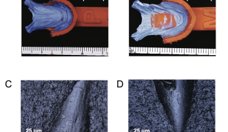
The impression process begins with sedating the animal. Next, the oral cavity is prepared by gently air-drying the teeth and using a high-viscosity putty impression material to clean them. This is immediately followed by the application of a more precise, low-viscosity polyvinyl siloxane material (an impression material widely used in dentistry) in custom-made, prefabricated 3D-printed trays to capture detailed impressions. The entire procedure typically takes 5 to 10 minutes.


One of the main challenges faced by the researchers was working with the small size of the fish, as their jaws were only about the size of a finger and individual teeth were less than a millimeter long. Other limitations included the need for precise cutting of the impressions for scanning and the inability to see inside the teeth structure. However, the researchers successfully performed the procedure on 60 fish with no fatalities. They observed detailed microwear patterns – tiny patterns in the tooth surface resulting from use over time.
Non-destructive tooth tracking
Dr. Ray Sallan, a dentist and researcher at OIST’s Science and Technology Group, described how the method provides several significant advantages over traditional techniques: “Previously, researchers had to euthanize specimens to study their teeth using CT scans or other methods. This new approach allows for non-destructive examination of living specimens, enabling researchers to track tooth replacement and development over time. It’s very valuable for studying rare species or museum specimens that can’t be damaged.”
The new technique has broad applications in various fields. It can be used to study microwear patterns to understand feeding habits, which is particularly useful in comparing modern species with fossils to determine ancient dietary patterns. The method can also be applied to study jaw biomechanics, track developmental changes, and examine comparative anatomy across species.
OIST PhD student and co-first author, Johannes Wibisana, from the Genomics and Regulatory Systems Unit highlighted the technique's versatility in studying different animals. "By checking the same features across different species, we can objectively compare variations due to diet, growth issues, or genetics. This method allows us to create plots showing differences between species or individuals. Dental traits from diverse species provide a valuable data set for analysis," he said.
The researchers are currently working on new experiments using this method with larger fish specimens and other vertebrates. They are particularly interested in studying tooth replacement patterns, which have never been quantified in living fish before. Only mammals have permanent adult teeth, while other vertebrates regularly grow new teeth throughout their lives.
"Our method has many potential applications and can be widely used, especially by museums and researchers sampling biodiversity. We can now safely and economically study and compare mouth structures, revealing differences and meticulous information that wasn’t previously accessible," Prof. Lauren Sallen, leader of the Macroevolution Unit and senior author, added.
Article Information
Specialties
Research Unit
For press enquiries:
Press Inquiry Form


