Okinawans at the Forefront
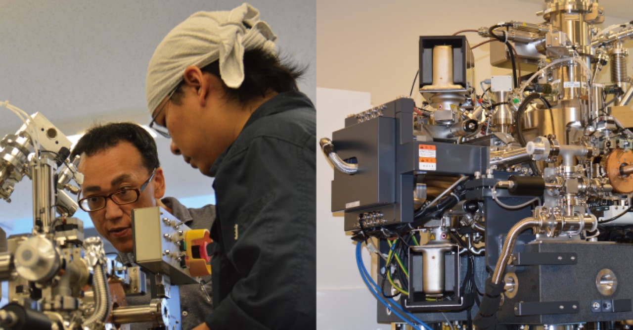
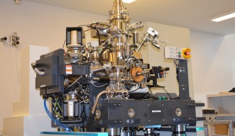
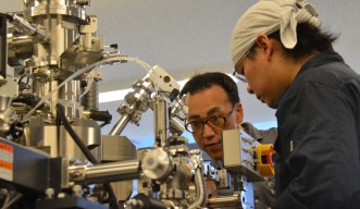
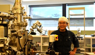
Professor Tsumoru Shintake’s Quantum Wave Microscopy Unit is developing a brand-new, world-class electron microscope which promises to make major contributions in biology and physics. The key players in the development of this microscope are two Okinawan researchers, Dr. Hidehito Adaniya and Dr. Ryo Yamashiro. “The reason why I had these two men come was because I thought they would do their best. They gave me the impression that they had perseverance and that they had guts,” Prof. Shintake said.
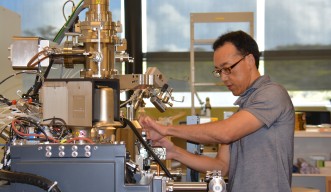
Dr. Yamashiro feels that having OIST, an international research institution, created more opportunities for local Okinawan researchers. Before coming to OIST a year ago, both Dr. Adaniya and Dr. Yamashiro pursued research outside of Okinawa. Dr. Adaniya worked at a research laboratory in the USA and investigated what happens to small molecules when the electron beams, which are used in electron microscopes to obtain the magnified image, interact with them. Dr. Yamashiro, on the other hand, investigated the dynamics of chemical reactions at the quantum level, and in doing so, used vacuum chambers like those that will contain the lens in the Quantum Wave Microscopy Unit’s electron microscope. Based on their previous experience, Dr. Adaniya carries out the simulations of the trajectory of electron beams, while Dr. Yamashiro designs the vacuum chamber. They work in a chilly room that is maintained at 23.5 – 24.5 degrees Celsius because the microscope is minutely designed and may malfunction if the temperature changes.
“The most important thing about developing a microscope is to be persistent,” says Prof. Shintake. Developing a microscope is an arduous process. Since metal parts are intricately assembled to make the whole microscope, even a 0.1mm miscalculation in the blueprint can prevent the machine from operating. Dr. Yamashiro sets himself to work with a sense of humor. “I really concentrate throughout the entire day to get the job done. I don’t take a moment to relax at work, but I’m fine because I go home and relax with my baby daughter.”
Once this microscope is complete, it will become much easier to observe biological samples. For instance, viruses have DNA enclosed within protein shells. Although most microscopes have permitted observation of viruses up to the shell structure level, this microscope will go further by enabling the imaging of the DNA. Another prospect is the detailed observation of nanoparticles such as carbon nanotubes.
This microscope will be fully assembled this October, and after a series of adjustments, the first prototype will be complete. “This microscope would have only been a dream if it were not for Prof. Shintake’s attitude towards research. He’s a researcher that really knows what it’s like at the research site. He can make radical ideas come to life because he visits the research site frequently and gets himself involved in the actual work,” says Dr. Adaniya. Same goes for the two Okinawan researchers; if it weren’t for their devotion, this innovative microscope would never have seen the light of day.
Specialty
Research Unit
For press enquiries:
Press Inquiry Form


