


mgu FY2013 Annual Report 3.1 fig1

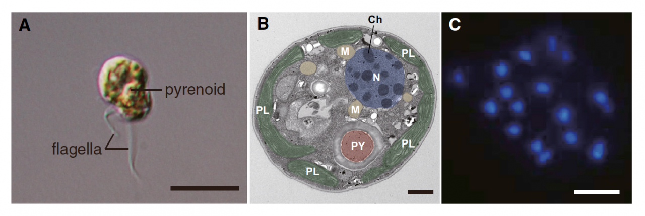
Figure 1. A Dinoflagellate, Symbiodinium minutum. (A) S. minutum zoospore. A short, transverse flagellum originating from the cingulum and a long, longitudinal flagellum originating from the sulcus are evident in the zoospore. A pyrenoid is also visible. The scale bar represents 10 mm. (B) Electron micrograph showing permanently condensed chromosomes (Ch) of S. minutum. The nucleus (N) is shown in purple, plastids (PL) in green, mitochondria (M) in orange, and pyrenoid (PY) in brown. The scale bar represents 1 mm. (C) DAPI staining of the nucleus showing permanently condensed chromosomes of S. minutum. The scale bar represents 1 mm.
Copyright OIST (Okinawa Institute of Science and Technology Graduate University, 沖縄科学技術大学院大学). Creative Commons Attribution 4.0 International License (CC BY 4.0).


