


onu FY2015 Annual Report 5

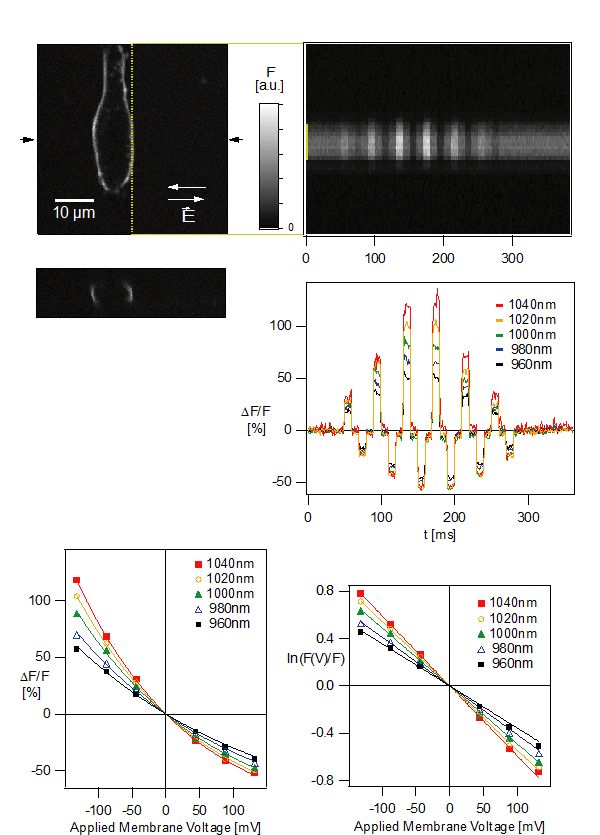
Figure: HEK293 cells labeled with ANNINE-6 are imaged with two-photon microscopy (top left). A line-scan is performed while alternating extracellular electric fields over the cell are applied (top right). The relative fluorescence change per applied voltage increases toward the spectral edge of the absorption spectrum of ANNINE-6 (bottom). (Kuhn et al 2004)
Date:
06 March 2024
Copyright OIST (Okinawa Institute of Science and Technology Graduate University, 沖縄科学技術大学院大学). Creative Commons Attribution 4.0 International License (CC BY 4.0).
Share on:


