


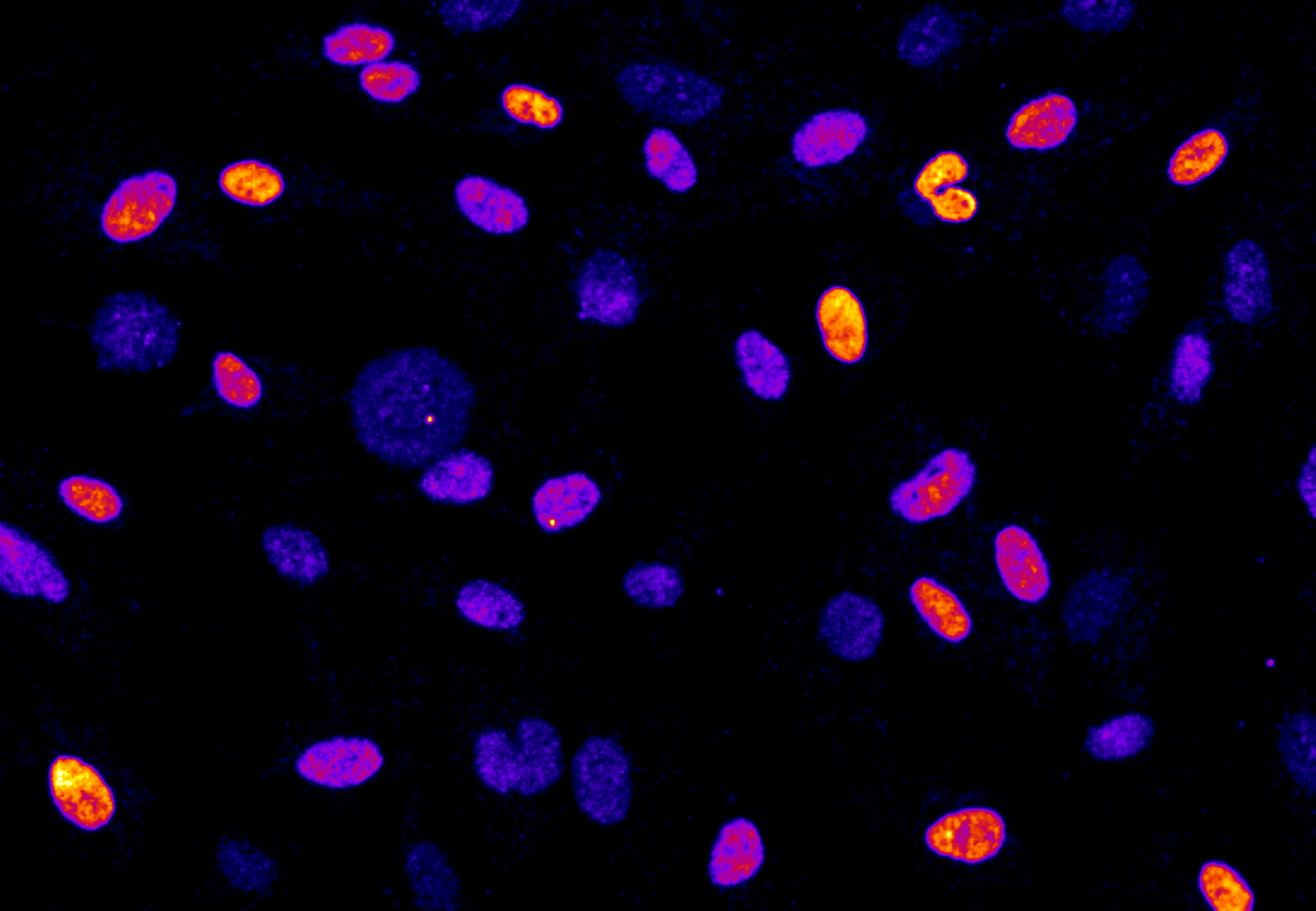
Cells under mitotic stress

The cells in this microscopy image have been stained with a fluorescent dye that binds only to proteins expressed in defective cells. This technique allows researchers to differentiate between cells that have experienced mitotic stress (yellow), and healthy cells (blue).
Date:
14 February 2024
Copyright OIST (Okinawa Institute of Science and Technology Graduate University, 沖縄科学技術大学院大学). Creative Commons Attribution 4.0 International License (CC BY 4.0).
Share on:


