onu FY2011 Annual Report 3

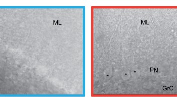
Figure 1:Astrocytes of the cerebellar cortex generate transglial calcium waves in vivo. (A) Bergmann glia cell expressing a fusion protein of the FCIP G-CaMP2 and DsRed under the CMV IE promoter after adenoviral gene transfer. (B) The use of FCIP also allows imaging of glial microdomains when cells are sparsely infected or (C) cell populations at higher infection rates which are almost comparable to (D) transgenic mice expressing GFP under the Bergmann glia-specific GFAP promoter. (E) Multicell bolus loading (MCBL) with the synthetic calcium dye fluo-5F/AM shows preferential Bergmann glia labeling if injected superficially into the molecular layer of the cerebellar cortex. (F) In vivo Bergmann glia show radially expanding transglial calcium waves measured with G-CaMP2. The first image shows resting fluorescence; following images show fluorescence changes relative to the first image. (G) Velate protoplasmic astrocyte of the granule cell layer expressing G-CaMP2 after infection by recombinant adenovirus. (H) Velate astrocytes also generate transglial calcium waves in vivo. The first image shows resting fluorescence; following images fluorescence changes relative to the first image. Scale bar in all panels, 20 µm.
Copyright OIST (Okinawa Institute of Science and Technology Graduate University, 沖縄科学技術大学院大学). Creative Commons Attribution 4.0 International License (CC BY 4.0).


