mbnu_F5 
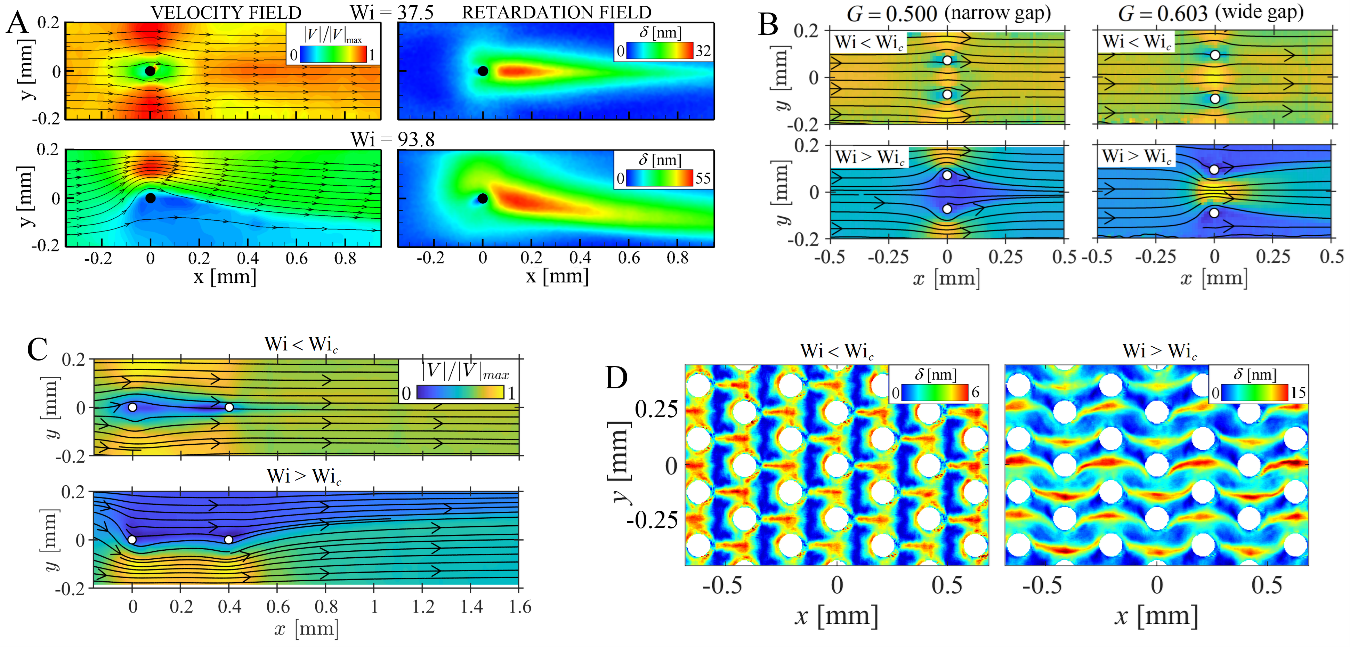
Figure 5: Transitions to steady asymmetric flow states in various geometries constructed from microscale cylinders as the Weissenberg number is increased beyond a critical value Wic. (a) Flow past a single cylinder positioned on the flow axis. (b) Velocity fields for flow past side-by-side cylinders with different dimensionless intercylinder gap, G = L1/(L1 + L2), where L1 and L2 are the cylinder-cylinder and cylinder-wall gaps, respectively. (c) Velocity fields for flow past two axially aligned cylinders. (d) Retardation fields for flow through a hexagonal array of cylinders. All cases show the flow from left to right of a shear-thinning viscoelastic WLM solution. Date: 25 March 2024 Copyright OIST (Okinawa Institute of Science and Technology Graduate University, 沖縄科学技術大学院大学). Creative Commons Attribution 4.0 International License (CC BY 4.0). Download full-resolution image Share on: Related Images Students from Gushikawa High School OIST aims to have every single high school student in Okinawa, like these students from Uruma City, visit the university before they graduate. ROV and xenophyophores A giant foraminifera called a xenophyophore appear as tiny dirt clods next to the robotic arm that collects them. Photo courtesy of JAMSTEC Xenophyophore An 8 cm large xenophyophore specimen from Ogasawara Trench showing clear growth rings. Photo courtesy of Beatrice Lecroq Warm or Cold? Brain slices of older tissues prepared with ice-cold (blue) and warm (red) preparation methods, as demonstrated by Shiwei Huang and Marylka Yoe Uusisaari in a paper in Frontiers in Cellular Neuroscience. The asterisks (*) indicate visible neurons. While no healthy neurons are visible in the left image, seven are visible in the right. Abbreviations: PN, Purkinje neuron layer; ML, molecular layer; GrC, granular cell layer. Shiwei Huang and Marylka Yoe Uusisaari Shiwei Huang (right) is a member of the Computational Neuroscience Unit and Marylka Yoe Uusisaari (left) is a part of the the Optical Neuroimaging Unit.
Students from Gushikawa High School OIST aims to have every single high school student in Okinawa, like these students from Uruma City, visit the university before they graduate.
ROV and xenophyophores A giant foraminifera called a xenophyophore appear as tiny dirt clods next to the robotic arm that collects them. Photo courtesy of JAMSTEC
Xenophyophore An 8 cm large xenophyophore specimen from Ogasawara Trench showing clear growth rings. Photo courtesy of Beatrice Lecroq
Warm or Cold? Brain slices of older tissues prepared with ice-cold (blue) and warm (red) preparation methods, as demonstrated by Shiwei Huang and Marylka Yoe Uusisaari in a paper in Frontiers in Cellular Neuroscience. The asterisks (*) indicate visible neurons. While no healthy neurons are visible in the left image, seven are visible in the right. Abbreviations: PN, Purkinje neuron layer; ML, molecular layer; GrC, granular cell layer.
Shiwei Huang and Marylka Yoe Uusisaari Shiwei Huang (right) is a member of the Computational Neuroscience Unit and Marylka Yoe Uusisaari (left) is a part of the the Optical Neuroimaging Unit.