


bsmu FY2016 Annual Report 04

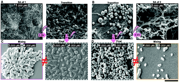
Figure 3: (A) SEM images of self-assemblies of 1, at 500 μM in H2O/DMSO (v/v = 3 : 2), and the transition state of photo-cleavage of SA of 1. (B) SEM images of self-assemblies of 2, at 500 μM in H2O/DMSO (v/v = 3 : 2), and the transition state of photo-cleavage of SA of 2. (C) SEM images of 3/4 mixture at 500 μM each in H2O/DMSO (v/v = 3 : 2) obtained from mixing or photo irradiation (320 nm) of SA of 1. (D) SEM images of 4/5 mixture at 500 μM each in H2O/DMSO (v/v = 3 : 2) obtained from mixing or photo irradiation (320 nm) of SA of 2. Scale bar of SEM is 1 μm.
Date:
01 March 2024
Copyright OIST (Okinawa Institute of Science and Technology Graduate University, 沖縄科学技術大学院大学). Creative Commons Attribution 4.0 International License (CC BY 4.0).
Share on:


