


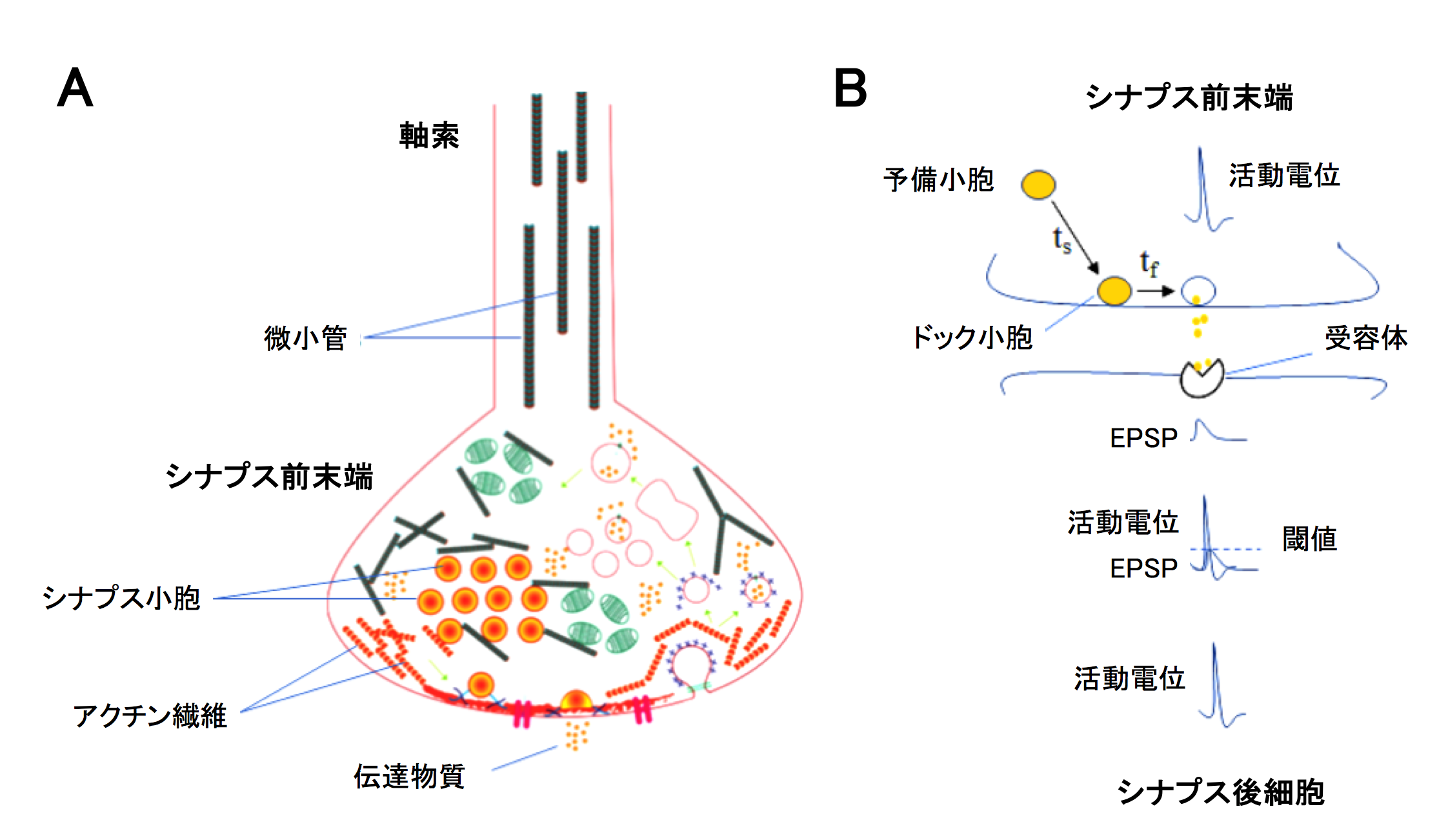
(A) Axon and presynaptic terminal. (B) Synaptic transmission.
Fig 1 (A) Axon and presynaptic terminal. (B) Synaptic transmission.
The thick filament “microtubules” (MTs) made of tubulin polymers and the thin filament “F-actin” made of actin polymers are important cytoskeletal elements supporting cell structures and functions. However, it remains unknown what role cytoskeletal proteins play in the neurotransmission process. In neurons, MTs are expressed in axons (Fig 1A) and serve as rails for transporting molecules and organelles between cell bodies and presynaptic terminals. In contrast, F-actin is expressed exclusively in presynaptic terminals. Presynaptic terminals contain “synaptic” vesicles (SVs) filled with neurotransmitter. When the electrical signal “action potentials” (APs) reach presynaptic terminals, SVs docked on the release sites fuse into terminal membrane, thereby releasing transmitters via exocytosis (Fig 1B). At the excitatory synapse, transmitter glutamate is released from SVs. Glutamate diffuses and binds to postsynaptic glutamate receptors and produces the postsynaptic response “EPSP”. When the EPSP size exceeds a threshold, APs are generated and propagate toward the axon terminal of postsynaptic neuron.
Copyright OIST (Okinawa Institute of Science and Technology Graduate University, 沖縄科学技術大学院大学). Creative Commons Attribution 4.0 International License (CC BY 4.0).
Tags


