


Zebrafish Retina Development

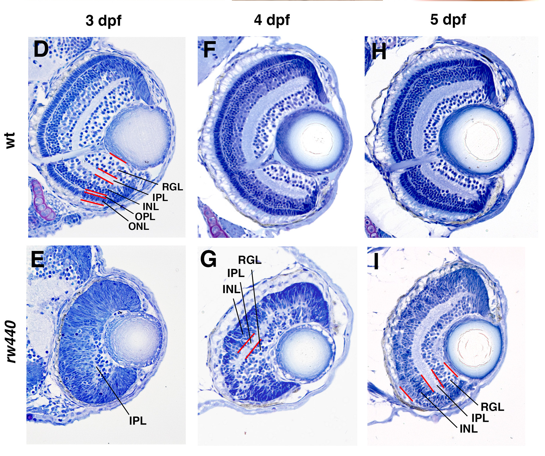
This figure shows the differences between a wild type zebrafish, labeled wt, and the SLBP mutant used in this experiment, labeled rw440. Moving from left to right, the images show the embryo at 3, 4, and 5 days post fertilization, or dpf. In the top row, the wild type fish retina forms different types of retinal cells, which assemble into neat layers. The wild type fish’s retinal ganglion cells form a clear channel leaving the eye for their axons to carry messages to the brain. On the bottom row, the mutant fish retina shows fewer types of cells, with delayed layering and no clear way for signals to reach the brain.
Copyright OIST (Okinawa Institute of Science and Technology Graduate University, 沖縄科学技術大学院大学). Creative Commons Attribution 4.0 International License (CC BY 4.0).
Tags


